Resources
Our team of communicators work hard to translate all the science behind AutoPiX and bring the power of imaging biomarkers closer to the non-expert community, including patients. We are constantly developing resources to help you speed-up with the AutoPiX findings including a glossary, webinars, and summaries of readings.
Readings
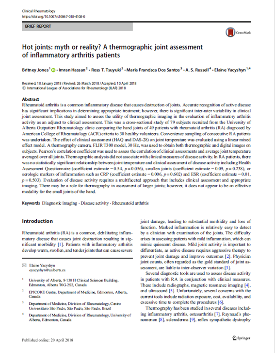
Hot joints: myth or reality? A thermographic joint assessment of inflammatory arthritis patients
Jones B, Hassan I, Tsuyuki RT, Dos Santos MF, Russell AS, Yacyshyn E. Hot joints: myth or reality? A thermographic joint assessment of inflammatory arthritis patients. Clin Rheumatol, 2018 Sep;37(9):2567-2571.
https://doi.org/10.1007/s10067-018-4108-0
The study examined whether thermographic imaging could be a useful tool for evaluating disease activity in rheumatoid arthritis (RA) patients. The researchers carried out thermography of hand joint temperatures of 49 RA patients compared with 30 healthy volunteers.
The results showed that joint temperatures were higher overall in RA patients compared to healthy volunteers. However, thermographic analysis did not correlate with clinical activity measures. No significant association was observed between joint temperature and Health Assessment Questionnaire scores, number of swollen joints, inflammatory markers (C-reactive protein and erythrocyte sedimentation rate) and Disease Activity Scores (DAS-28). In healthy volunteers, right hands were significantly warmer than left hands, regardless of hand dominance.
The researchers concluded that thermography does not appear to be an effective modality for assessing inflammation in the small joints of the hand in RA patients. However, it might have applications for larger joints. The authors suggest that evaluation of disease activity requires a multifaceted approach that includes clinical assessment and appropriate imaging.
Why is this important for AutoPiX?
The detailed protocol of this study to standardise imaging (controlled room temperature, resting periods, fixed camera distances) provides valuable methodological insights for conducting imaging studies in arthritis. Besides, the article offers critical insights into the limitations of thermography for small joint assessment in RA and emphasises that disease activity evaluation requires a multifaceted approach. Alternative imaging techniques (radiographs, MRI, ultrasound) help to contextualise where thermography fits within the broader landscape of arthritis imaging that AutoPiX may be exploring. Negative findings, like those observed in this study, are crucial for advancing the field by narrowing down which imaging approaches deserve further investigation for arthritis assessment.
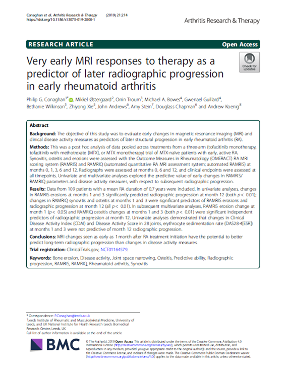
Very early MRI responses to therapy as a predictor of later radiographic progression in early rheumatoid arthritis
Conaghan PG, Østergaard M, Troum O, Bowes MA, Guillard G, Wilkinson B, Xie Z, Andrews J, Stein A, Chapman D, Koenig A. Very early MRI responses to therapy as a predictor of later radiographic progression in early rheumatoid arthritis. Arthritis Res Ther, 2019 Oct 21;21(1):214.
https://doi.org/10.1186/s13075-019-2000-1
The purpose of this article is to evaluate whether early MRI changes and clinical disease activity measures can predict later structural progression in early RA patients included in a clinical trial. MRI assessments were conducted at multiple time points to measure synovitis, osteitis, and erosions using RAMRIS and RAMRIQ scoring systems.
The results showed that early MRI changes, particularly in RAMRIS erosions and RAMRIQ osteitis and synovitis at months 1 and 3, were significant predictors of radiographic progression at month 12. On the contrary, changes in clinical disease activity measures (CDAI and DAS28-4[ESR]) at months 1 and 3 did not predict radiographic progression at month 12.
These results suggest the potential of MRI as a tool for predicting disease progression and guiding personalised treatment strategies in RA. Incorporating early MRI measurements in clinical trials could enable more rapid assessment of treatment efficacy and potentially inform treatment decisions in clinical practice.
Why is this important for AutoPiX?
Overall, the insights from this article underscore the potential of imaging in enhancing the management of arthritis, which is central to the objectives of the AutoPiX project.
The ability to predict long-term outcomes based on early imaging data can support clinicians in making informed treatment decisions. AutoPiX can develop tools, such as early MRI biomarkers, that integrate these predictive insights into clinical practice. This approach can contribute to personalised treatment strategies and timely adjustments in therapy, potentially improving patient outcomes.
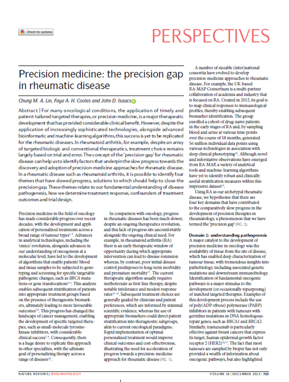
Precision medicine: The precision gap in rheumatic disease
Lin CMA, Cooles FAH, Isaacs JD. Precision medicine: the precision gap in rheumatic disease. Nat Rev Rheumatol, 2022 Dec;18(12):725-733.
https://doi.org/10.1038/s41584-022-00845-w
The article examines the disparity between the advancements in precision medicine in oncology and the relatively slower progress in rheumatic diseases, particularly rheumatoid arthritis (RA).
This "precision gap" can be explained by different factors. Unlike oncology, where tissue biopsies have provided deep insights into disease mechanisms, RA research has been limited by a lack of synovial biopsies and a poor understanding of disease pathobiology. This has hindered the identification of robust biomarkers for targeted therapies.
In RA, outcome measures like DAS28 are composite and subjective, making it challenging to link treatment responses to specific biological changes. Molecular and cellular outcome measures may be more effective in reflecting disease biology. RA clinical trials often include patients with established disease and multiple comorbidities, which can confound treatment outcomes. Factors such as disease duration, previous therapies, and demographic variables need to be carefully considered in trial design and analysis. The authors advocate for more efficient trial designs.
To close the precision gap, the authors point to the need for a better understanding of RA pathobiology, improved outcome measures, and more sophisticated trial designs. It also emphasises the importance of collaborative efforts among researchers, clinicians, and regulators to advance precision medicine in rheumatic diseases.
Why is this important for AutoPiX?
This article highlights the importance of precision medicine in improving patient outcomes. By leveraging advanced imaging techniques, AutoPiX can contribute to the development of personalised treatment strategies for arthritis patients.
Imaging biomarkers or imaging-based outcome measures developed through the AutoPiX project, which will be tested against biopsies, could provide objective measures of disease activity and progression. These biomarkers could be used to stratify patients in clinical trials, ensuring more efficient and precise results. In all, the AutoPiX project can play a crucial role in the collaborative effort called for in this article by providing innovative imaging solutions that enhance patient care and research outcomes.
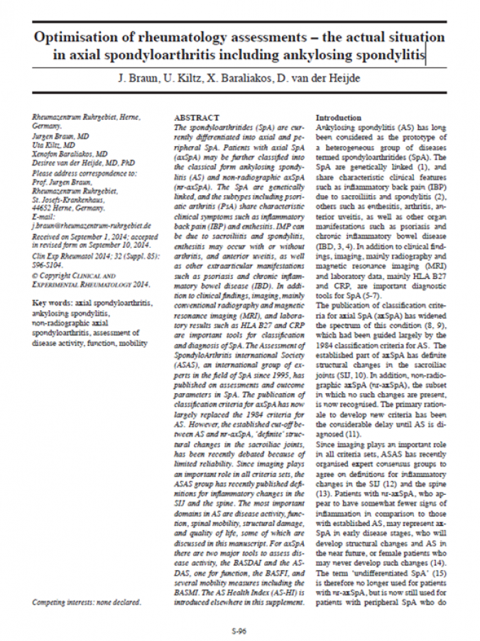
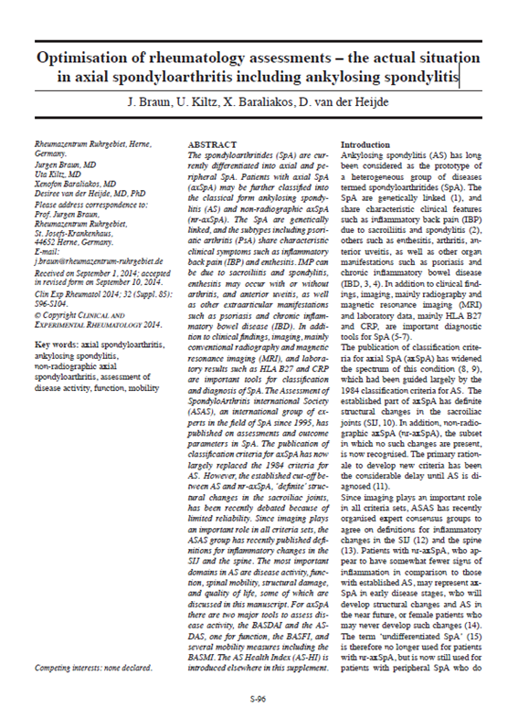
Optimisation of rheumatology assessments – the actual situation in axial spondylarthritis, including ankylosing spondylitis
Braun J, Kiltz U, Baraliakos X, van der Heijde D. Optimisation of rheumatology assessments - the actual situation in axial spondyloarthritis including ankylosing spondylitis. Clin Exp Rheumatol, 2014 Sep-Oct;32(5 Suppl 85):S-96-104
This article provides an overview of the current state of assessments in axial spondyloarthritis (axSpA), including ankylosing spondylitis (AS). It emphasises the importance of comprehensive assessments in axSpA, highlighting the strengths and limitations of current tools.
Axial spondyloarthritis is divided into AS and non-radiographic axSpA (nr-axSpA). Diagnosis involves clinical findings, imaging (radiography and MRI), and laboratory results (HLA B27 and C-reactive protein, CRP).
Main disease activity measures include BASDAI (Bath Ankylosing Spondylitis Disease Activity Index), a patient-reported measure that assesses fatigue, spinal pain, joint pain/swelling, areas of localized tenderness, and morning stiffness; and ASDAS (Ankylosing Spondylitis Disease Activity Score), a composite index that includes patient-reported measures, CRP or ESR, and provides a more comprehensive assessment of disease activity. ASDAS is recommended for its superior performance in measuring disease activity and guiding treatment decisions.
Functional assessment is based on BASFI (Bath Ankylosing Spondylitis Functional Index), which measures functional ability through 10 items scored by the patient and the ICF (International Classification of Functioning, Disability, and Health).
Spinal mobility can be assessed by the BASMI (Bath Ankylosing Spondylitis Metrology Index) with age-specific reference intervals to guide clinicians. Imaging, particularly MRI, is crucial for diagnosing and monitoring axSpA. Disease activity measures, such as ASDAS, are associated with radiographic progression and can guide treatment decisions.
Why is this important for AutoPiX?
This article highlights the critical role of imaging, particularly MRI, in the diagnosis and monitoring of axSpA. It underlines the need to focus on the development of more comprehensive and patient-centred assessment tools.
Imaging can contribute significantly to the more comprehensive assessments by providing objective data on disease progression and activity. The insights from this article can help the AutoPiX project in developing imaging solutions that are clinically relevant, patient-centred, and tailored to individual patient needs. By understanding the current landscape of disease assessment in axSpA, the AutoPiX project can better integrate its imaging solutions into existing clinical workflows, ensuring that they complement current practices.
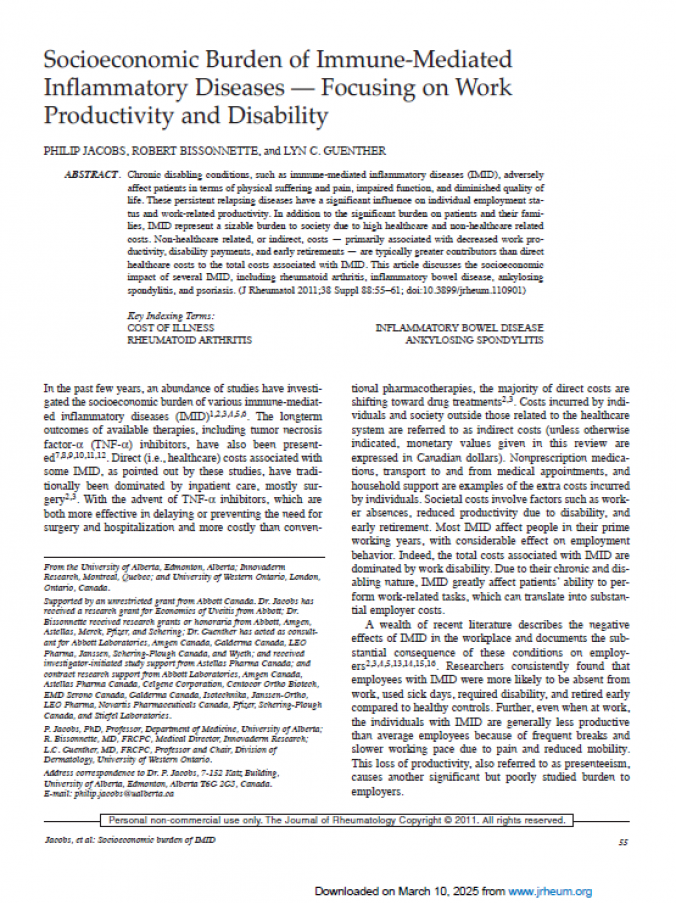
Socioeconomic burden of immune-mediated inflammatory diseases. Focusing on work productivity and disability
Jacobs P, Bissonnette R, Guenther LC. Socioeconomic burden of immune-mediated inflammatory diseases--focusing on work productivity and disability. J Rheumatol Suppl. 2011 Nov;88:55-61.
https://doi.org/10.3899/jrheum.110901
Chronic disabling conditions, such as immune-mediated inflammatory diseases (IMID), adversely affect patients in terms of physical suffering and pain, impaired function, and diminished quality of life. These persistent relapsing diseases have a significant influence on individual employment status and work-related productivity. In addition to the significant burden on patients and their families, IMID represents a sizable burden to society due to high healthcare and non-healthcare-related costs. Non-healthcare related, or indirect, costs — primarily associated with decreased work productivity, disability payments, and early retirements — are typically greater contributors than direct healthcare costs to the total costs related to IMID. Direct or healthcare costs associated with IMID have traditionally been dominated by inpatient care, mostly surgery. With the advent of therapies that are more effective in delaying or preventing the need for surgery and hospitalisation, the majority of direct costs are shifting toward drug treatments. Costs incurred by individuals and society outside those related to the healthcare system are referred to as indirect costs. Non-prescription medications, transport to and from medical appointments, and household support are examples of the extra costs incurred by individuals. Societal costs involve factors such as worker absences, reduced productivity due to disability and early retirement. Most IMIDs affect people in their prime working years, with a considerable effect on employment behaviour. Indeed, the total costs associated with IMID are dominated by work disability. Socioeconomic outcomes, including work productivity, are an essential piece of the puzzle when conducting economic evaluations. Evidence regarding their scope and magnitude should be included in economic assessments, along with direct costs and health outcomes. Adverse effects of IMID concerning socioeconomic burden and work productivity have been well documented. The emergence of biologic therapies has revolutionised the treatment of these disabling diseases and made long-lasting remission highly achievable.
Why is this important for AutoPiX?
The study highlights the socioeconomic burden of IMID, including arthritis and its impact on work productivity and disability. Understanding this impact can help justify the need for advanced imaging techniques that improve diagnosis and management, potentially reducing the overall burden on patients and society. By incorporating these outcomes into the development and evaluation of imaging techniques, the AutoPiX project can ensure that its innovations address the broader needs of patients and that its imaging solutions are developed and evaluated with a holistic view of patient and societal benefits.
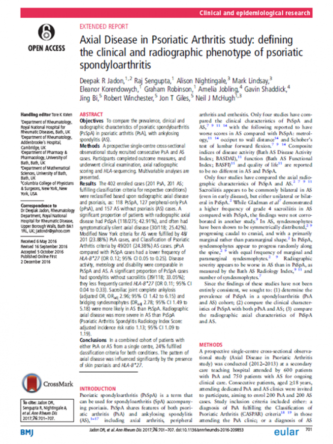
Axial disease in psoriatic arthritis study: Defining the clinical and radiographic phenotype of psoriatic spondylarthritis
Jadon DR, Sengupta R, Nightingale A, Lindsay M, Korendowych E, Robinson G, et al. Axial Disease in Psoriatic Arthritis study: defining the clinical and radiographic phenotype of psoriatic spondyloarthritis. Ann Rheum Dis, 2017 Apr;76(4):701-707.
https://doi.org/10.1136/annrheumdis-2016-209853
Psoriatic spondylarthritis (PsSpA) describes the presence of spondylarthritis (SpA) accompanying psoriasis. PsSpA shares features of both psoriatic arthritis (PsA) and ankylosing spondylitis (AS), including axial arthritis, peripheral arthritis and enthesitis. The results of studies comparing the clinical characteristics of PsSpA and AS have not been entirely consistent, suggesting a high degree of overlap between them. This article aims to define the similarities and differences between these conditions and whether clinical and genetic parameters could explain these. Axial disease was defined radiographically. Accordingly, a significant proportion of PsA cases were reclassified to the PsSpA phenotype and found to be often asymptomatic. Notably, a quarter of PsA and AS cases were classifiable to the other disease using the currently available classification systems. In PsSpA cases, spondylitis without sacroiliitis was common and usually symptomatic. Previous studies have shown a similarly high frequency of spondylitis without sacroiliitis and proposed it as a feature distinguishing PsSpA. AS cases were more likely than PsSpA cases to have complete sacroiliac joint ankylosis and bridging syndesmophytes. These features may prove more useful than syndesmophyte shape to distinguish PsSpA from AS. The results of this study indicate that PsSpA forms part of the SpA spectrum. In terms of most clinical indices of disease activity, PsSpA has a similar disease burden as AS, despite being less severe radiographically. This may be of relevance in the development of future treatment guidelines.
Why is this important for AutoPiX?
Understanding the overlap and differences between PsSpA and AS is crucial for developing imaging protocols that accurately distinguish between these conditions. The article emphasises the importance of radiographic definitions in classifying axial disease in PsA and AS and highlights that imaging could play a vital role in identifying subclinical cases. The article provides valuable clinical and radiographic insights that can inform the development and application of imaging techniques in the AutoPiX project, ultimately contributing to better patient outcomes in arthritis management.
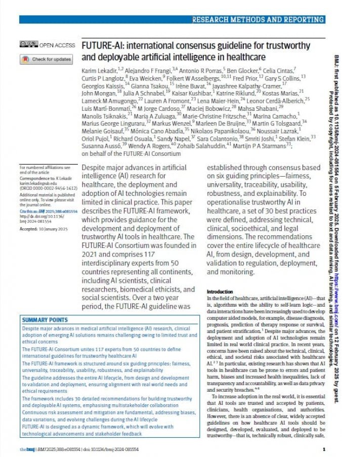
FUTURE-AI: international consensus guideline for trustworthy and deployable artificial intelligence in healthcare
Lekadir K et al. BMJ 2025; 388:e081554. http://dx.doi.org/10.1136/bmj‑2024‑081554
Despite significant advancements in AI research for healthcare, the actual use of these technologies in clinical practice remains limited, mainly due to concerns about trust, ethics, and practical deployment. To help solve this, over 100 experts worldwide came together to create FUTURE-AI, a set of international guidelines to ensure AI tools in healthcare are trustworthy, safe, and practical. The FUTURE-AI framework is built around six guiding principles:
- Fairness: AI should perform equally well across different groups of people, avoiding biases related to gender, age, ethnicity, etc.
- Universality: AI tools should be generalisable and work well in various clinical settings and populations.
- Traceability: There should be mechanisms to document and monitor the entire lifecycle of AI tools to ensure transparency and accountability.
- Usability: AI tools should be user-friendly and integrate smoothly into clinical workflows, enhancing productivity and patient outcomes.
- Robustness: AI tools should maintain performance under real-world variations and unexpected conditions.
- Explainability: AI decisions should be understandable to clinicians and patients, and they should provide clear explanations for their outputs.
The guideline was developed using a modified Delphi approach, involving multiple rounds of feedback and consensus-building among experts and includes 30 recommendations covering the entire lifecycle of healthcare AI, from design and development to validation, deployment, and monitoring. The FUTURE-AI guideline is designed to be dynamic, evolving with technological advancements and stakeholder feedback to ensure its relevance and effectiveness.
Why is this important for AutoPiX?
The FUTURE-AI framework is highly relevant to the AutoPiX project for several reasons:
- Guidelines for Trustworthy AI: For the AutoPiX project, which likely involves AI applications in healthcare, adhering to these guidelines can ensure that the AI tools developed are reliable, ethical, and effective.
- Ethical and Fair AI: AutoPiX can benefit from these principles to ensure its AI tools do not perpetuate biases and are equitable across different patient groups.
- Robustness and Usability: AutoPiX can leverage these guidelines to create user-friendly AI solutions that are resilient to real-world variations, ensuring seamless integration into clinical workflows.
- Explainability and Transparency: For AutoPiX, ensuring that AI decisions are transparent and understandable to clinicians and patients can enhance trust and facilitate better clinical decision-making.
- Regulatory Compliance: AutoPiX can use these guidelines to navigate the complex regulatory landscape and ensure that its AI tools meet necessary legal and safety standards.
- Stakeholder Engagement: For AutoPiX, involving healthcare professionals, ethicists, and patients in the development process can lead to AI tools more aligned with real-world needs and challenges.
- Dynamic and Evolving Framework: This adaptability benefits AutoPiX, as it allows the project to stay current with best practices and emerging standards in AI development.
By aligning with the FUTURE-AI framework, the AutoPiX project can enhance its credibility, ensure ethical AI deployment, and improve the likelihood of successful integration and adoption in clinical settings.
Neural networks for automatic scoring of arthritis disease activity on ultrasound images
Andersen et al. RMD Open 2019;5:e000891. doi:10.1136/rmdopen-2018-000891
This article explores the use of convolutional neural networks (CNNs), a type of AI model (See Glossary), to automate the scoring of disease activity in rheumatoid arthritis (RA) using Doppler ultrasound (US) images according to the OMERACT-EULAR Synovitis Scoring (OESS) system, aiming to reduce interobserver variability and enhance objectivity in clinical practice and trials.
The study utilised two CNN architectures to classify US images from RA patients. One network classified the images as healthy or diseased, while the other scored images across all four OESS categories. The networks were trained on a dataset of 1694 images and tested on a separate set. The results showed that CNN, classifying images as healthy or diseased, achieved accuracies of 86.4% and 86.9%, with high sensitivity and specificity. The CNN for full-scale OESS scoring achieved an average accuracy of 75% and strong agreement with expert rheumatologist scores.
These results demonstrated that CNNs can effectively evaluate joint US images, potentially providing a more unbiased method for scoring US arthritis activity. This technology could enhance consistency in clinical assessments and trials, particularly in settings with limited access to musculoskeletal imaging experts. The authors suggest further optimisation of CNN designs, including combining classifiers from different network layers and expanding the dataset to include more joints, which could improve performance and applicability in clinical settings.
Why is this important for AutoPiX?
This research highlights the potential of AI in enhancing the objectivity and efficiency of disease activity assessments in rheumatology. The insights from this article support the development of imaging technologies that enhance the objectivity, consistency, and efficiency of arthritis assessments, which are central goals of the AutoPiX project. This consistency is crucial for accurate diagnosis, treatment planning, and monitoring disease progression. Automated scoring can help manage the workload in clinical settings, especially with a shortage of specialists. This efficiency can lead to faster and more accurate assessments, benefiting patient care.
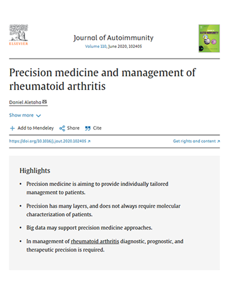
Precision medicine and management of rheumatoid arthritis
Aletaha D. J
Autoimmun 2020 Jun:110:102405.
Doi: 10.1016/j.jaut.2020.102405
Exploring the relevance of precision medicine in rheumatoid arthritis for the AutoPiX project, highlighting the integration of AI and big data for personalized treatment approaches.
Precision medicine (PM) is a commonly used term that implies a highly individualized and tailored approach to patient management. PM implies using the unique molecular characteristics of a patient for management decisions. This discipline has moved from using information of an individual patient to selecting the best existing therapy to target discovery by identifying subgroups of patients with specific characteristics, for which targeted therapies can possibly be developed. In clinical practice profiling patients is only useful, if this subsequently can allow targeting the patient’s disease more precisely therapeutically. Examples of precision medicine from oncology may define the potential for other diseases such as rheumatoid arthritis. Oncologists already use therapy with autologous T cells which have a genetically engineered chimeric antigen receptor (so called, CAR-T cells) that can specifically target and kill respective tumor cells. These compounds are precise in individual patients but at the same time, are highly costly; therefore, adequate patient selection clearly needs to be scrutinized. One consequence of precision medicine approaches is a growing need for genetic counseling, and providers are lining up to allow the identification of the right patient for these approaches.
Big data and precision medicine are terms that commonly go together. Today, the presence of large-scale biologic databases and powerful platforms for characterizing patients have created an incredible amount of potentially available information from each patient. At the same time the computation tools for analyzing this amount of data have expanded dramatically.
In addition to molecular sources, big data can also come from other sources (environment, sociodemographic, nutrition) and technologies (electronic health record, mobile phone applications, and wearables). Once obtained, the next step is to integrate these heterogeneous data bioinformatically. The integrated “big data” can then be analyzed using different approaches like machine learning, or network based and statistical methods.
The four major areas for precision medicine in RA include diagnosis, prognosis, treatment selection and treatment reduction. Precision medicine and big data are a fast-evolving field which, despite the current limitations regarding practical relevance, will likely change rheumatology in the years to come.
The convergence of precision medicine and AI in rheumatoid arthritis enhances personalized care and aligns with AutoPiX's mission to improve diagnostic and therapeutic outcomes.
Why is this important for AutoPiX?
The article by Aletaha D. is important for the AutoPiX project because it highlights the potential of precision medicine in managing rheumatoid arthritis (RA), emphasizing personalized treatment approaches based on individual patient characteristics. This aligns with AutoPiX's goal of using AI to enhance imaging biomarkers for RA, facilitating more precise diagnosis and treatment decisions. The article also discusses the role of big data and computational tools like machine learning in precision medicine, which is similar to how AutoPiX leverages AI to analyze imaging data and convert it into meaningful biomarkers. Both the article and the AutoPiX project focus on addressing critical unmet needs in rheumatology, such as improving access to advanced imaging techniques and selecting appropriate treatments. By enhancing imaging biomarkers, AutoPiX contributes to the future of rheumatology, where precision medicine is expected to play a significant role, as suggested by the article
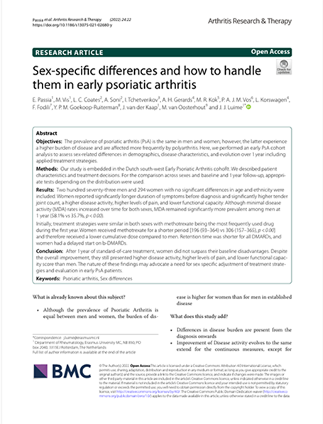
Sex-specific differences and how to handle them in early psoriatic arthritis
E Passia, M Vis, L C Coates, A Soni, I Tchetverikov, A H Gerards et al.
Arthritis Research & Therapy 2022; 24:22.
Doi: 10.1186/s13075-021-02680-y
This study highlights significant sex-specific differences in the clinical manifestations and treatment outcomes of early psoriatic arthritis, suggesting a need for tailored treatment approaches.
Sex and health is a new area of study, aiming to investigate the differences between men and women in both health an disease. Sex has been shown to affect natural history, clinical manifestations, and response to medications in several rheumatic diseases. Although the prevalence of psoriatic arthritis (PsA) is considered equal in men and women, the are not equally affected, with women experiencing a higher burden of disease (pain, disability and fatigue).
The objective of this paper is to assess sex-related differences in patients with newly diagnosed PsA and evaluate the evolution over the first year, in terms of disease activity and health-related quality of life.
The study was conducted on 620 patients (307 men and 313 women). There were no differences in the age at onset of PsA but women reported longer duration of symptoms before diagnosis, less frequency of paid employment and higher prevalence of obesity. At baseline, women presented higher tender joint counts, more frequency of axial disease and worse results in composited indices than men. Fatigue, anxiety and comorbid medical conditions were also more common in women who suffered more severe limitations in function and worse quality of life compared with men. On the contrary, skin lesions were more common in men. At 1 year of follow-up men remained in minimal disease activity and remission more frequently than women. Despite the improvement presented by both sex through 1 year follow-up, women reported higher levels of pain and worse functional status. Cumulative doses of specific drugs were lower in women despite the higher disease burden observed, may be explained by a more frequent reporting of side effects compared to men.
Overall, women presented higher disease activity, pain, and functional impairment compared to men at baseline but also at 1 year of follow-up. Women seem to be undertreated. The nature of these findings could suggest that sex bias in prescribing exists and may advocate the need for sex specific adjustment of treatment strategies and evaluation of Psa.
The study underscores the importance of considering sex-specific differences in the management of psoriatic arthritis, as women experience a higher disease burden and may be undertreated due to differences in symptom reporting and treatment tolerance.
Why is this important for AutoPiX?
The findings from this study on sex-specific differences in early psoriatic arthritis are highly relevant to the AutoPix project, as they emphasize the need for personalized and gender-sensitive approaches in disease management. By understanding these differences, AutoPix can develop more effective diagnostic and therapeutic strategies that account for the unique experiences of both men and women with psoriatic arthritis, potentially improving treatment outcomes and quality of life for all patients. This could involve integrating sex-specific data into AI-driven diagnostic tools and treatment planning algorithms to ensure more tailored care.
Distinguishing rheumatoid arthritis from psoriatic arthritis
Merola JF, Espinoza LR, Fleischmann R. Distinguishing rheumatoid arthritis from psoriatic arthritis.
RMD Open 2018;4:e000656.
doi:10.1136/rmdopen-2018-000656
Differentiating between RA and PsA can be challenging because both diseases present many similarities.
Rheumatoid arthritis (RA) and psoriatic arthritis (PsA) are autoimmune systemic inflammatory diseases characterised by pain and swelling in the joints with systemic manifestations. Joint involvement is predominantly symmetric in RA and often asymmetric in PsA. Most patients have polyarthritis (≥5 involved joints), although the involvement can be oligo or polyarticular. The involvement of the axial skeleton in PsA can be a differentiating feature because it is not present in RA other than cervical spine involvement. Enthesitis, dactylitis and nail dystrophy are common in PsA. Ocular disorders present as keratoconjunctivitis sicca in RA, and as uveitis in PsA. Cutaneous manifestations are common in RA (rheumatoid nodules) and PsA (psoriasis). RA is seropositive for RF or CCP antibodies and PsA is seronegative. Comorbidities linked to systemic inflammation (e.g. cardiovascular disease) are common in both, although the burden may be higher in RA. Obesity, diabetes and metabolic syndrome are significantly higher in PsA.
Although the pathogenesis of RA and PsA is not entirely understood, different factors are thought to trigger an autoimmune inflammatory response. In both RA and PsA, inflammatory responses are characterised by the increased production of proinflammatory molecules associated with joint damage, radiographic progression, and gut inflammation, but different pathways are involved.
Because of the differences in pathogenesis, clinical manifestations, and response to therapy between RA and PsA, treatment strategies may differ.
Why is this important for AutoPiX?
The key differentiating features between RA and PsA—such as the presence of psoriasis, joint involvement patterns, and specific biomarkers (e.g., seropositivity for RA vs. seronegativity for PsA)—could be incorporated into an AI diagnostic system that automatically identifies the type of arthritis based on patient data.
